Case history – 45 year old person with multiple cervical lymph nodes.
This series of case of the week is for Postgraduates in pathology – to stay updated with amazing hand picked cases from practice.
If you wish to contribute to case of the week – https://pathocubs.com/contact-us/ – Your credentials will be added to the post. let’s make this a one stop solution for all the pathology needs.
Every month – Winners of 4 case of the weeks will be identified and will be awarded with prizes ! Participate and let’s make pathology learning wonderful !


Non hodgkin lymphoma s/o DLBCL
Burkitt’s lymphoma
Burkitt lymphoma
Non hodgkins lymphoma
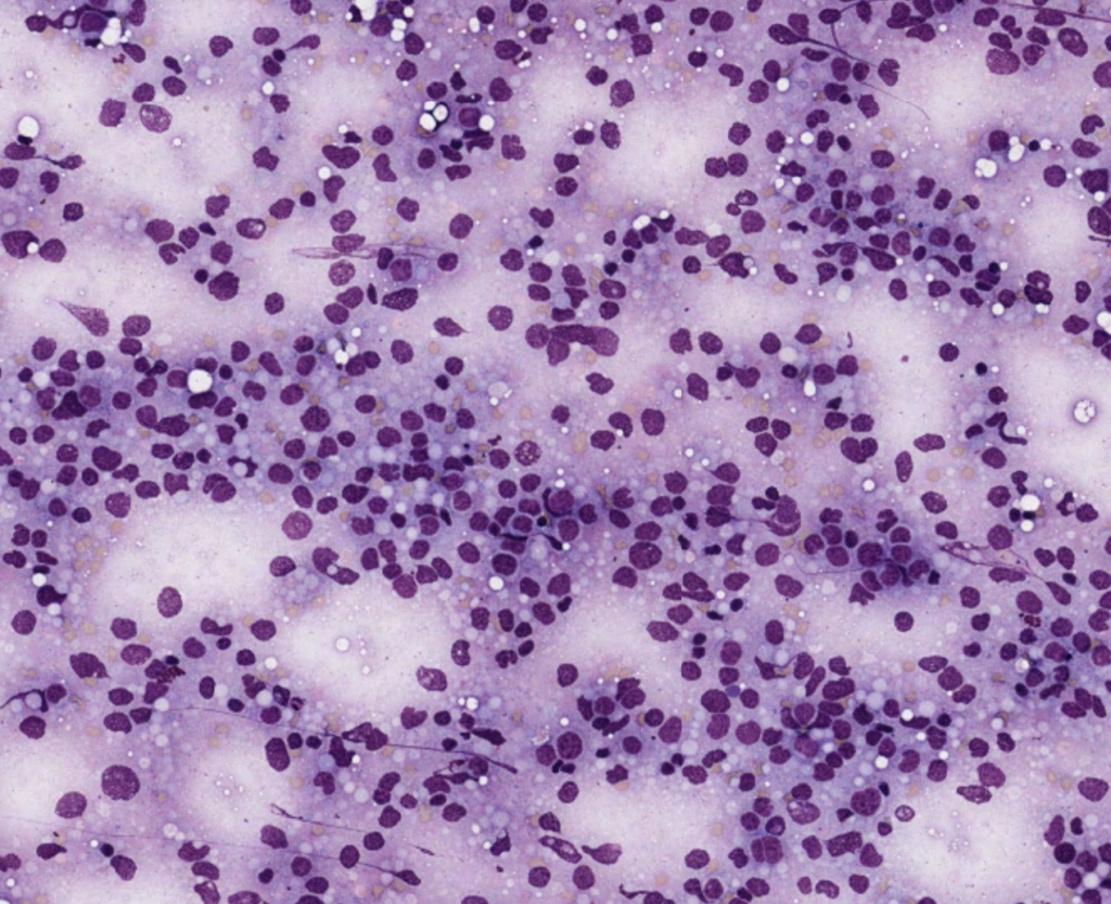
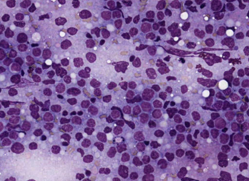
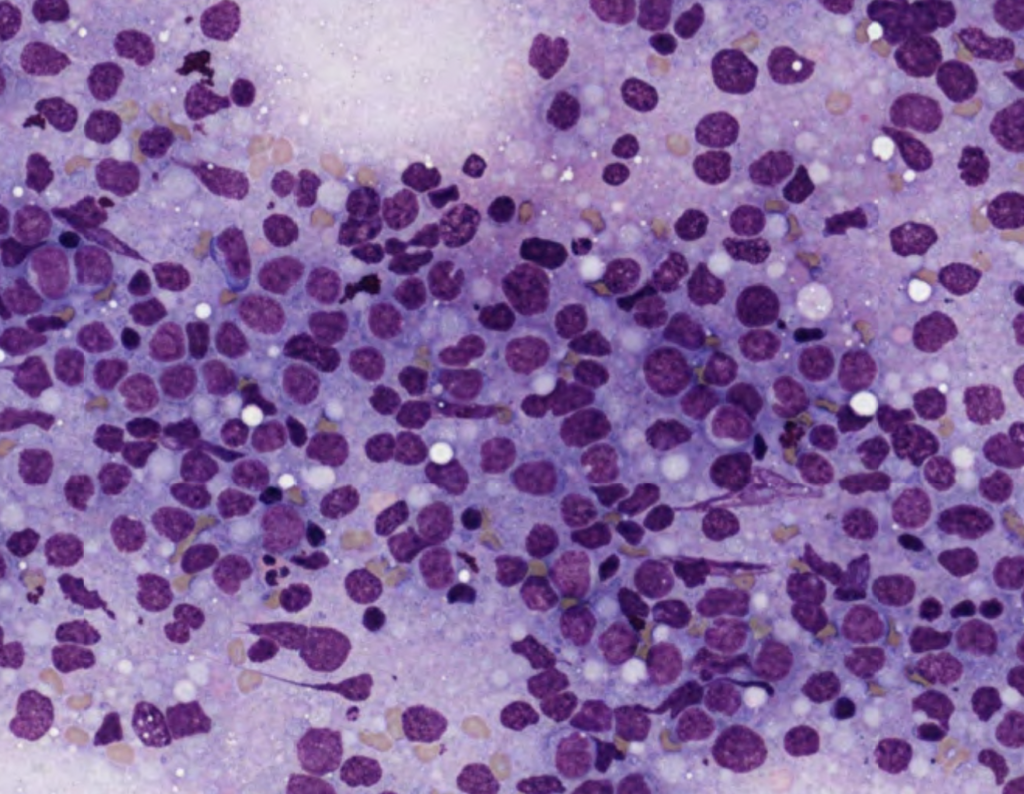
Aspirated smears are highly cellular showing sheets of atypical cells which are medium to large size, with high N:C ratio, and scant cytoplasm. Few showing prominent nucleoli. Background shows mature lymphocytes admixed with RBCs
IMPRESSION – FNAC – Highly cellular smears – suggestive of lymphoproliferative disease cytologically favouring Non Hodgkins lymhphoma
Non Hogkins Lymphoma with Medium to large sizes cells. D/D : Anaplastic large cell lymphoma
Diffuse large B cell lymphoma
Sorry Medium to small to medium-sized lymphoid cells.
D/d : Follicular lymphoma
CLL/SLL
Mantle cell lymphoma
Marginal zone lymphoma
DLBCL
NHL
Hypercellular Smear studied shows polymorphous population of cells comprised of lymphocytes,histiocytes,plasma cells,eosinophils,arranged in diffuse solid sheetsin a hemorrhagic nodal background.There are centrocytes centroblast,follicular dendritic cells,.Also seen few large cells,binucleated with pr nucleoli
Possibilities to be considered
1. Hodgkins lymphoma
2. Mets from undiff ca (sinonasal)
Features in favor of Non-Hodgkin’s lymphoma
Advice excision biopsy for further subtyping
Hypercellular smear
Monotonous neoplastic cells
Round to oval nuclei with basophilic cytoplasm, containing lipid vacuoles
Impression: Burkitts lymphoma.
Non hodgkin’s lymphoma- probably diffuse large B cell lymphoma
Hyper cellular smears showing diffuse infiltration of large sized lymphoid cells with round to oval nuclei, coarse chromatin, several nucleoli and moderate amount of blue cytoplasm. Apoptotic bodies seen.
Impression : NHL
Cellular smears show atypical cells arranged in sheets. Cells are medium to large with round to oval nucleus, irregular nuclear contour, increased N:C ratio, and scant basophilic cytoplasm.Background shows blood elements and few mature looking lymphocytes.
The above cytomorphological features are suggestive of a lymphoproliferative disorder(Non Hodgkins lymphoma)
D/D Follicular lymphoma
Adv:Clinicoradiological correlation &
HPE for confirmation
Cellular smear shows a polymorphic population of cells arranged in sheets. The cells are small to large, comprising cells of lymphoid origin with few cells having cleaved nucleus and others having a round to oval nucleus with inconspicuous nucleoli. Mild to moderate nuclear pleomorphism is noted in the cells. There is lymphocytic infiltration in the background.
The above cytomorphological features goes in favour of Non Hodgkin Lymphoma probably Follicular Lymphoma.
Aspirated smears are highly cellular showing sheets of atypical cells which are intermediate sized with round to oval nuclei ,high N:C ratio, coarse chromatin and moderate vacuolated cytoplasm intermixed with scanty small round lymphocytes and histiocytes and . Few showing prominent nucleoli. Also seen are few mitotic figures.
Features are in favour of Non Hodgkin lymphoma- D/D 1. Burkitt lymphoma
2. Mantle cell lymphoma
IHC: Bcl6 and bcl 2for confirmation
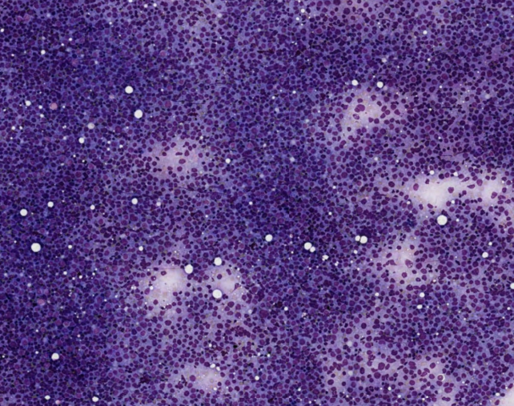
Approach; Low power showing small royal blue cytoplasm, with pale areas giving a starry sky appearance.
cytosmears studied shows rich cell yield consisting of atypical lymphocytes arranged in sheets,clusters which are 2-3 times increased in size compared to normal mature lymphocytes having increased nuclear cytoplasmic ratio,irregular clumping of chromatin admixed with macrophages and normal looking lymphocytes against necrotic debri in hemorrhagic background.
Impression: LYMPHOPROLIFERATIVE DISEASE-NON HODGKIN LYMPHOMA
NOTE: Advised lymph node biopsy for Histopathological confirmation
Cytosmears are cellular showing small to medium sized atypical lymphoid cells admixed with mature lymphocytes and histiocytes. The atypical cells have high N:C ratio, irregular nuclear membrane, scant cytoplasm and conspicuous nucleoli.
Features are suggestive of lymphoma.
Following possibilities can be considered:- 1. NHL
2. Follicular lymphoma
Highly cellular smear shows heterogeneous population of medium to slightly enlarged atypical cells with irregular nuclei scant to moderate amount of cytoplasm irregular nuclear membrane granular chromatin prominent 1-2 nuclei and intranuclear inclusions admixed with few lymphocytes, plasma cells suggestive of Non hodgkins lymphoma DD marginal zone lymphoma
Follicular lymphoma
Cellular smear showing lymphoid cells arranged in sheets cells are medium to large sized with inconspicuous nucleoli and scant cytoplasm.
Diagnosis: lymphoproliferative disorder non hodgkins lymphoma
Suggest cell block with ihc : cd5, cd20, bcl2, cd10, bcl6 , mum1 and cyclind1
Cellular smears study show ,discretely arranged cells having scant cytoplasm with high nuclear to cytoplasmic ratio with coarse chromatin with few of them having nuclear indentations,in a background of small mature lymphocytes .
features suggestive of lymphoproliferative disorder probably Non Hodgkin lymphoma.
S/o NHL probably DLBCL